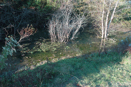
Once again the microaquarium appears to be full of life.
The diatoms are still high in numbers. The Tachysomas seem to have reduced in number.


The organism pictured above was identified as Anacystis cyanea (Forest 1954) This organism was of particular interest and I had a good time trying to identify it with some assistance from Dr. McFarland. It has a blue-green color and a rounded cluster. It remained stationary.
Another organism was identified as Centropyxis sp. (Patterson 2003) It was an amoeba with a shell that it made around itself. the shell had an opening and you could see the amoeba moving around inside. There were multiples of this organism located around the microaquarium. They are stationary.
This was the last observation for the microaquarium lab.
Bibliography
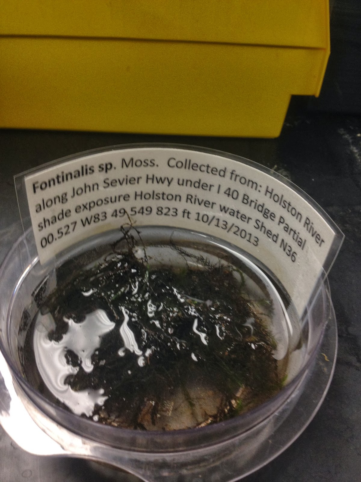
Patterson, D.J. Free-Living Freshwater Protozoa: A Colour Guide. Washington, D.C. Mason Publishing. 2003. Pg 95
Forest, H.S. Handbook of Algae. The University of Tennessee Press. 1954. Pg 366.
Prescott, G.W. How to Know The Fresh-Water Algae. WM. C. Brown Company Publishers. 1964. Pg 187.